Table of Contents
Loose connective tissue also announced as Areolar connective tissue is a class of connective tissue that includes areolar tissue, reticular tissue, and adipose tissue. Areolar connective tissue is the most familiar type of connective tissue in vertebrates. It carries organs in place and attaches epithelial tissue to other underlying tissues. For example, it creates telae, such as the tela submucosa and tela subserosa, which attach mucous and serous membranes to the muscular layer. It also besieges the blood vessels and nerves.
Cells hailed fibroblasts are broadly circulated in this tissue; they are intermittent branching cells that secrete strong fibrous proteins and proteoglycans as an extracellular matrix. The cells of this type of tissue are generally detached by quite some distance by a gelatinous substance mainly made up of collagenous and elastic fibers. Where is areolar tissue absent Usually “areolar connective tissue” is examined as a parent category that consists of the mucous connective tissue of the fetus, areolar connective tissue, reticular connective tissue, and adipose tissue.
Areolar Connective Tissue
Areolar connective tissue is the most common CT of the body. It is characterized by plenty of ground substance, plus thin and comparably few fibers and cells. The main cellular elements are fibroblasts and a more miniature amount of adipocytes. Fat cells are an essential constituent of areolar connective tissue, but when they are bountiful and coordinated into large lobules for storage purposes the tissue is better evaluated as adipose tissue.
The adipocytes present in areolar connective tissue are generally isolated cells or small aggregations that do not function as storage depots, and their principal function is to clarify gliding and to act as interstitial fillers. The adipocytes of the areolar connective tissue generally do not increase in volume when entities gain weight. Collagen is the dominant fiber of areolar connective tissue and is displayed in all directions to form a loose network in the intercellular material. Many elastic fibers are also present.
The areolar combinative tissue has a viscous, gel-like consistency and its consistency may fluctuate in disparate parts of the body due to variations in temperature or pH. This CT allows descending between the various muscles and organs and empowers the diffusion of oxygen/nutrients from small vessels to the cells and the dissemination of metabolites back to the vessels. It is the initial site where antigens, bacteria, and other agents that have breached an epithelial surface can be damaged.
It also develops a mesh-like tissue with a fluid matrix that bolsters the epithelia, such as the skin and other membranes. This CT fills the spaces between disparate organs and thus holds them in place while softening and protecting them; it also surrounds and reinforces the blood vessels.
A particular category of areolar connective tissue is the reticular tissue that consists of only reticular fibers formed of type III collagen. The reticular cells have a stellate shape and long processes that make contact with neighboring cells, and the subsequent tissue holds a number of bodily structures, such as the liver, spleen, bone marrow, and lymphatic organs.
The Structure Of Areolar Tissue
Areolar Tissue is the areolar connective tissue that consists of a meshwork of collagen, elastic tissue, and reticular fibers – with many connective tissue cells in between the meshwork of fibers.
The fibers that construct the mesh structure of areolar tissue include:
- Collagen Fibres
- Elastic Fibres
- Reticular Fibres
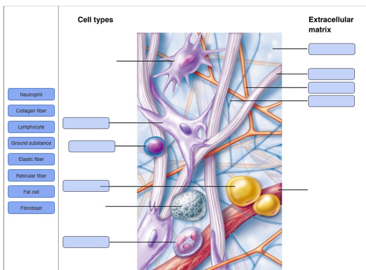
The disparate categories of cells embedded within the areolar tissue include:
- Fibroblasts
- Plasma Cells
- Adipocytes
- Mast Cells
- Macrophages
All of the above (fibers and cells) are buried in a semi-fluid ground substance – which is identical to the ground substance in which the chondrocytes are established in hyaline cartilage tissue.
Location
It may be found in tissue sections from almost every part of the body. It surrounds blood vessels and nerves and cracks with them even into the small spaces of muscles, tendons, and other tissues. It may likewise be present in the mediastinal extremities.
Nearly every epithelium rests on a slab of areolar tissue, whose blood vessels contribute the epithelium with nutrition, waste elimination, and a ready supply of infection-fighting leukocytes when needed. Because of the abundance of open, fluid-filled space, leukocytes can advance about effortlessly in areolar tissue and can easily find and destroy pathogens.
The areolar tissue is found beneath the epidermis layer and is also underneath the epithelial tissue of all the body arrangements that have external openings. it makes the skin elastic and benefits it to withstand pulling pain. It is also a constituent of the lamina propria of the digestive and respiratory tracts, the mucous sheaths of reproductive and urinary systems, the stroma of glands, and the hypodermis of the skin. It is also found in the mesentery which is surrounding the intestine.
Fibers
Areolar connective tissue is named depended on the “weave” and type of its constituent fibers. There are three main types:
- Collagenous fibers: collagenous fibers are made of collagen and consist of bundles of fibrils that are braids of collagen molecules.
- Elastic fibers: elastic fibers are made of elastin and are “stretchable.”
- Reticular fibers: reticular fibers consist of one or more types of very thin collagen fibers. They join connective tissues to other tissues.
Function
Areolar connective tissue arrests organs in place and attaches epithelial tissue to other underlying tissues. It also serves as a reservoir of water and salts for surrounding tissues. Almost all cells obtain their nutrients from and release their wastes into areolar connective tissue.
Areolar Connective Tissue:
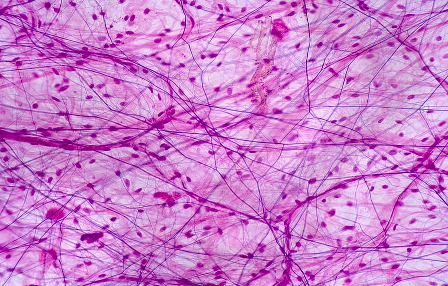
Areolar connective tissue is a type of connective tissue that is widely distributed throughout the body. It is composed of a loosely arranged network of collagen, elastic fibers, and cells called fibroblasts. The term “areolar” refers to the tissue’s sponge-like appearance, with small spaces or gaps between the components. This tissue provides support, elasticity, and flexibility to various organs and structures.
Loose Areolar Connective Tissue:
Loose areolar connective tissue is a subtype of areolar connective tissue characterized by its loose arrangement of fibers and cells. It forms a delicate, flexible framework within organs and helps to hold them in place. This type of tissue is found in spaces between muscles, around blood vessels and nerves, and beneath the epithelial layers of organs. It also serves as a cushioning material between organs.
Areolar Tissue Location:
Areolar tissue is found in various locations throughout the body, including the skin, mucous membranes, and organs. In the skin, it is located in the dermis, the layer beneath the epidermis. Areolar tissue is also present in the submucosa of the digestive tract, the subcutaneous layer beneath the skin, and the lamina propria of various organs, such as the respiratory tract and urinary bladder.
Areolar Connective Tissue Location:
Areolar connective tissue is located in similar areas as areolar tissue. It can be found beneath the skin, surrounding blood vessels and nerves, in the submucosa of organs, and between muscles. This tissue acts as a supportive and binding material, filling spaces and providing structural integrity.
Areolar Tissue Function:
The primary functions of areolar tissue include providing support, connecting different tissues and organs, and facilitating the exchange of nutrients and waste products. It acts as a universal packing material, filling the spaces between organs and providing structural support. Areolar tissue also plays a crucial role in the immune response, as it contains various immune cells that defend against pathogens.
Areolar Tissue Anatomy:
Areolar tissue has a complex anatomy. It consists of a network of collagen and elastic fibers that form a three-dimensional framework. Fibroblasts are the predominant cells within this tissue and are responsible for producing and maintaining the extracellular matrix. Other cells, such as macrophages and mast cells, are also present and contribute to the tissue’s functions.
Areolar Tissue Location and Function:
The location and function of areolar tissue are closely intertwined. Its location in various organs and structures allow it to fulfill its functions effectively. For example, in the skin, areolar tissue provides elasticity and support to the dermis, allowing the skin to stretch and recoil. In organs like the digestive tract, areolar tissue helps to anchor the mucous membranes and provides flexibility during movement.
Areolar Tissue Example:
An example of areolar tissue can be found in the subcutaneous layer beneath the skin. This layer consists of a loose arrangement of collagen and elastic fibers, as well as fibroblasts. It serves as a thermal insulator, protecting the body from temperature changes, and acts as a cushioning material, protecting underlying structures from external forces.
Areolar Tissue Types:
Areolar tissue is considered a type of loose connective tissue. Other types of loose connective tissue include adipose tissue (fat tissue) and reticular tissue. Adipose tissue stores energy in the form of fat, while reticular tissue forms a supporting network within certain organs, such as the liver and spleen. These different types of loose connective tissue have distinct characteristics and functions.
In summary, areolar tissue is a versatile connective tissue that is widely distributed throughout the body. Its loose arrangement of fibers and cells allows it to provide support, flexibility, and protection to various organs and structures. Understanding the location, function, and anatomy of areolar tissue is crucial for comprehending its role in maintaining the integrity and functionality of the body.
Frequently Asked Questions About Areolar Connective Tissue
Where Is Areolar Tissue Found And What Is Its Function?
Located In The Skin, Areolar Tissue Binds The Outer Layers Of The Skin To The Muscles Lying Underneath. They Are Also Found In, Around The Mucous Membranes, Surrounding Nerves, Blood Vessels And Various Other Body Organs. Its Functions Are As Follows: Supports The Internal Organs.
What Are The Main Functions Of Areolar Connective Tissue?
These Tissues Are Widely Distributed And Serve As A Universal Packing Material Between Other Tissues. The Functions Of Areolar Connective Tissue Include The Support And Binding Of Other Tissues. It Also Helps In Defending Against Infection.
What Is An Example Of Areolar Connective Tissue?
The Areolar Tissue Is A Loose Connective Tissue That Can Be Seen Between The Skin And Muscles; In The Bone Marrow As Well As Around The Blood Vessels And Nerves. These Adipocyte Cells Together Make The Adipose Tissue Or The Fat Tissue.
What Are The Characteristics Of Areolar Connective Tissue?
This Is A Loose Connective Tissue Widely Spread Throughout The Body. It Contains All Three Types Of Fibers (Collagen, Elastin, And Reticular) With Much Ground Substance And Fibroblasts.
Where Is Areolar Tissue Found?
Areolar tissue is found in various locations throughout the body. It forms a widespread network that fills the spaces between organs, muscles, and other tissues. Some specific locations where areolar tissue can be found include:
– Beneath the skin (subcutaneous layer): Areolar tissue provides a supportive and cushioning layer beneath the skin.
– Surrounding blood vessels and nerves: Areolar tissue surrounds and supports blood vessels and nerves throughout the body.
– Submucosa of organs: Areolar tissue is present in the submucosal layer of organs such as the digestive tract, respiratory tract, and urinary bladder.
– Lamina propria: Areolar tissue forms the connective tissue layer known as the lamina propria beneath the epithelial lining of various mucous membranes.
Where Is Areolar Connective Tissue Found In The Body?
Areolar connective tissue is found in similar locations to areolar tissue. It is a widely distributed connective tissue that can be found throughout the body, including:
– Subcutaneous layer: Areolar connective tissue is present beneath the skin, providing support and insulation.
– Surrounding blood vessels and nerves: It surrounds and protects blood vessels and nerves, helping to anchor them in place.
– Submucosa of organs: Areolar connective tissue is found in the submucosal layer of organs like the digestive tract and respiratory tract, providing structural support.
– Lamina propria: It forms the lamina propria beneath the epithelial lining of various mucous membranes, such as those in the respiratory system, digestive system, and urinary system.
What Are The Visible Characteristics Of Areolar Connective Tissue?
Areolar connective tissue exhibits several visible characteristics:
– Loosely arranged fibers: The collagen and elastic fibers in areolar connective tissue are arranged in a loose, random manner, creating a spongy appearance.
– Abundance of cells: Areolar connective tissue contains various cells, primarily fibroblasts, which are responsible for producing and maintaining the extracellular matrix.
– Spaces or gaps: Areolar connective tissue has open spaces or gaps between its components, which allow for flexibility, nutrient exchange, and the presence of immune cells.
– Vascularized: This tissue is highly vascular, meaning it is well-supplied with blood vessels, ensuring the delivery of oxygen and nutrients.
Which Membrane Is Composed Of Areolar Connective Tissue And Not An Epithelial Tissue?
The membrane composed of areolar connective tissue and not an epithelial tissue is the synovial membrane. The synovial membrane lines the cavities of synovial joints, which are the freely movable joints in the body (e.g., knee joint, elbow joint). Unlike other membranes in the body that consist of an epithelial layer, the synovial membrane is composed of connective tissue, specifically areolar connective tissue.
The synovial membrane functions to secrete synovial fluid, which lubricates the joint and reduces friction between the articulating surfaces. It also nourishes the cartilage and cushions the joint during movement.
What Is An Example Of An Areolar Tissue?
One example of areolar tissue is the subcutaneous layer, also known as the hypodermis. The subcutaneous layer is located beneath the skin and is composed of areolar connective tissue. It serves as a supportive layer, providing insulation, cushioning, and protection to the underlying structures. The subcutaneous layer also contains adipose tissue (fat cells) that store energy and contribute to insulation.
The areolar tissue in the subcutaneous layer helps to anchor the skin to the underlying structures while allowing for movement and flexibility. It also contains blood vessels and nerves that supply the skin and regulate temperature.
Why Is It Called Areolar?
Areolar tissue is named after its appearance, which resembles an areola, the circular area of pigmented skin surrounding the nipple. The term “areolar” means “little open space” or “small area with clear boundaries.” This name is derived from the tissue’s structure, which consists of loosely arranged fibers and cells with visible spaces or gaps between them.
The areolar tissue’s open spaces allow for the diffusion of nutrients, gases, and other substances, as well as the migration of immune cells and the flexibility required for organ movement. The term “areolar” helps describe this characteristic appearance and arrangement of the tissue.