Table of Contents
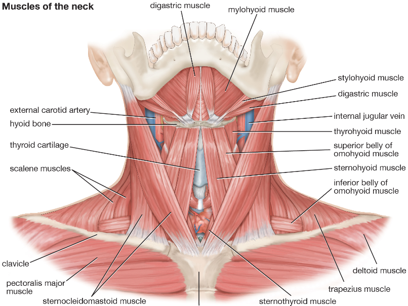
The digastric muscle (also digastricus) (named digastric as it has two ‘bellies’) is a small muscle located under the jaw. The term “digastric muscle” refers to this specific muscle. However, other muscles that have two separate muscle bellies include the ligament of Treitz, omohyoid, occipitofrontalis. It lies below the body of the mandible, and extends, in a curved form, from the mastoid notch to the symphysis menti. It belongs to the suprahyoid muscle group. A broad aponeurotic layer is given off from the tendon of the digastricus on either side, to be attached to the body and greater cornu of the hyoid bone; this is termed the suprahyoid aponeurosis.
The digastric muscle consists of the anterior belly and the posterior belly connecting the mandible, hyoid bone, and temporal bone. Its unique morphology, structure, and variations have drawn genuine interest in this muscle from anatomists, scientists, and physicians for a long time, and the variations of the digastric muscle have been documented since the 18th century. As the usage of computed tomography and magnetic resonance imaging in the neck has become ever increasing, recognizing the variations of the digastric muscle can be a great value since it helps physicians to make better treatment plans and avoid unnecessary invasive procedures in the neck. Although the variations of the digastric muscle do not necessarily cause clinical symptoms, they still have important clinical applications. This article discusses the anatomy, embryology, descriptions of the morphological variations, and clinical significance of the digastric muscle.
Digastric Muscle
The digastricus (digastric muscle) consists of two muscular bellies united by an intermediate rounded tendon. The two bellies of the digastric muscle have different embryological origins and are supplied by different cranial nerves. Each person has a right and left digastric muscle. In most anatomical discussions, the singular is used to refer to a muscle, even when each person actually has two of that muscle one on the right side, and another on the left. For example, we speak of the deltoid, even though there is one deltoid in each shoulder. Likewise, we speak of the digastric even though there is a right and left digastric muscle.
Posterior belly
The posterior belly, longer than the anterior belly, arises from the mastoid notch which is on the inferior surface of the skull, medial to the mastoid process of the temporal bone. The mastoid notch is a deep groove between the mastoid process and the styloid process. The mastoid notch is also referred to as the digastric groove or the digastric fossa. The posterior belly is supplied by the digastric branch of the facial nerve. The digastric muscle stretches between the mastoid process of the cranium to the mandible at the chin, and part-way between, it becomes a tendon that passes through a tendinous pulley attached to the hyoid bone. It originates from the second pharyngeal arch.
Anterior belly
The anterior belly arises from a depression on the inner side of the lower border of the mandible called the Digastric fossa of Mandible, close to the symphysis, and passes downward and backward. The anterior body is supplied by the trigeminal via the mylohyoid nerve, a branch of the inferior alveolar nerve, itself a branch of the mandibular division of the trigeminal nerve. It originates from the first pharyngeal arch.
Intermediate tendon
The two bellies end in an intermediate tendon which perforates the Stylohyoideus muscle and is held in connection with the side of the body and the greater cornu of the hyoid bone by a fibrous loop, which is sometimes lined by a mucous sheath.
Variations
Variations are numerous.
The posterior belly may arise partly or entirely from the styloid process, or be connected by a slip to the middle or inferior constrictor; the anterior belly maybe double or extra slips from this belly may pass to the jaw or Mylohyoideus or decussate with a similar slip-on opposite side; anterior belly may be absent and posterior belly inserted into the middle of the jaw or hyoid bone. The tendon may pass in front, more rarely behind the Stylohoideus. The Mentohyoideus muscle passes from the body of the hyoid bone to the chin.
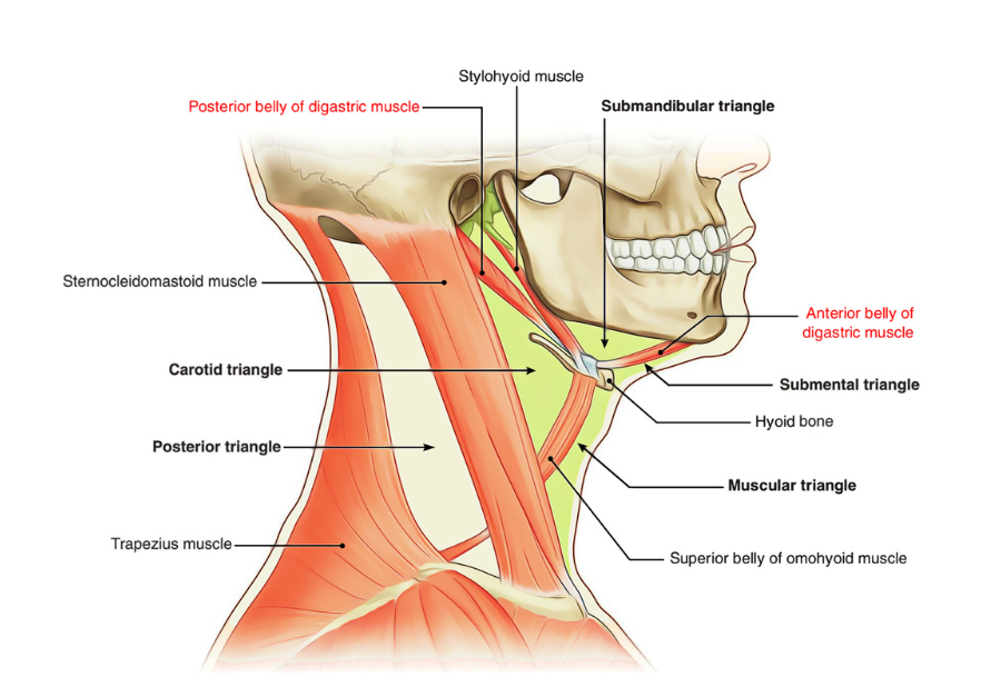
Triangles
The Digastricus divides the anterior triangle of the neck into three smaller triangles.
- the submandibular triangle(also called Digastric Triangle), bounded above by the lower border of the body of the mandible, and a line drawn from its angle to the Sternocleidomastoideus, below by the posterior belly of the Digastricus and the Stylohyoideus, in front by the anterior belly of the Diagastricus;
- the carotid triangle, bounded above by the posterior belly of the Digastricus and Stylohyoideus, behind by the Sternocleidomastoideus, below by the Omohyoideus;
- the suprahyoid or submental triangle, bounded laterally by the anterior belly of the Digastricus, medially by the middle line of the neck from the hyoid bone to the symphysis menti, and inferiorly by the body of the hyoid bone.
- The inferior carotid triangle (or muscular triangle), is bounded, in front, by the median line of the neck from the hyoid bone to the sternum; behind, by the anterior margin of the Sternocleidomastoideus; above, by the superior belly of the Omohyoideus
The function of the Digastric Muscle
Unlike most muscles, the digastric muscle can contract in two different portions. Because the bellies of the muscle are supplied by different nerves, the muscles can contract separately. While the exact workings of the face and jaw muscles are very complicated, the digastric muscle works as a kind of tension pulley to generate forces in different directions on the jaw. There are two digastric muscles, one on the left and right, which attach to the lower skull through a pulley mechanism at the hyoid bone. This means that the contraction of this muscle can produce forces that open the jaw and wiggle it from side-to-side. The digastric muscle is thus responsible for actions such as speaking, chewing, swallowing, and breathing. Any complex action of the jaw will likely involve the muscle in some way.
Digastric Muscle Pain
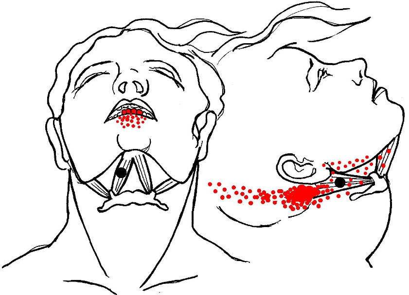
The digastric muscle is often pinpointed as the source in people experiencing jaw, throat, tooth, and general facial pain. The muscle, having two portions separately innervated by different branches of cranial nerves and operating a complex tendon pulley, is prone to tense up. Unfortunately, the tension in the anterior belly of the muscle and tension in the posterior belly do not produce the same sensation. The posterior belly is connected to the facial nerve. The anterior belly is connected to the trigeminal nerve, which connects to different parts of the face and jaw than the facial nerve. Thus, any tension in either part of the muscle can send pain impulses through many parts of the face and jaw. Relaxing the muscle with some simple jaw movements and light massage should ease the tension and reduce the pain, even if it feels like it is not coming from the digastric muscle.
What Causes Digastric Muscle Pain?
The Upper Portions Of The Sternocleidomastoid Muscle Will Be Tender To The Touch A Result Of Trigger Points From The Posterior Belly Of The Digastric Muscle. The Digastric Can Also Cause A Deep Ear Pain Described As Being In Front Of Or Below The Ear That Is Not Caused By An Ear Infection.
What Is The Function Of The Digastric?
Lesson Summary
The Middle Of The Digastric Muscle, Which Is Where A Tendon Connects The Anterior And Posterior Bellies, Attaches To The Hyoid Bone. There Are Two Main Functions Of The Digastric Muscle: Depression Of The Mandible (Lower Jaw): Causes The Mouth/Jaw To Open. Elevation Of The Hyoid Bone: Aids In Swallowing.
What Does The Sternohyoid Muscle Do?
The Sternohyoid Muscle’s Main Function Is The Depression Of The Hyoid Bone. The Hyoid Bone Is Located Below The Mandible, Or Lower Jaw, And Is A ‘u’ Shaped Bone That Is Partially Responsible For Tongue Movement And The Action Of Swallowing. The Sternohyoid Is One Of A Pair Of Muscles Responsible For This Action.
What Is The Muscle Under The Chin?
The Transversus Menti, Or Transverse Muscle Of The Chin, Is A Facial Muscle That Is Often Considered To Be The Superficial Fibers Of The Depressor Anguli Oris Muscle Which Cross To The Other Side Of The Face.
How Do You Stop A Jaw Cramp?
Over-the-counter Pain Relievers: Medicines Like Ibuprofen And Acetaminophen May Help To Reduce Discomfort. Massage The Affected Joint: Using Your Index And Middle Finger, Press The Sore Areas Of Your Jaw, Such As The Area Right Before Your Ear Where Your Jaw Joints Meet.